This lecture is entitled, test your sonography skills, the importance of this lecture is to illustrate how ultrasounds can be used for clinical decision making. We are going to look at one particular case and see how the use of the ultrasound can influence your decision making.
Now please be aware that there are many different ways to approach what is considered a complex case here and by no means am I saying that this is the only way to approach the case, again the purpose is to show how an ultrasound or in this case, more than one ultrasound, can influence clinical decision making, so we will start off with the beginning of the case.
This is a sixty four year old male, septic from a urinary tract infection who has DIC and became short of breath requiring intubation, his base line chest X-ray and EKG were normal, his vital signs are remarkable for hypotension, tachycardia, and being specialist at thirty eight degrees, he is sedated from medications and in no acute distress, his lungs are remarkable for crackles, his heart has a systolic ejection murmur, his abdomen has decreased bowel sounds and is non tender.
This is a sixty four year old male, septic from a urinary tract infection who has DIC and became short of breath requiring intubation, his base line chest X-ray and EKG were normal, his vital signs are remarkable for hypotension, tachycardia, and being specialist at thirty eight degrees, he is sedated from medications and in no acute distress, his lungs are remarkable for crackles, his heart has a systolic ejection murmur, his abdomen has decreased bowel sounds and is non tender.
Bearing in mind however that he is sedated from medications making exams somewhat difficult, extremities are within normal limits and likewise for his neurological exam, there is no focal deficits but he is very sedated from medications and again the complete exam is somewhat difficult, the following tests were ordered, a chest X-ray was ordered, was performed on a stat basis and showed moderate congestive heart failure, an EKG was ordered and as soon as the EKG tech came to perform it, the patient was on the way to CT angiography and it was felt that that should be given priority so the EKG is still pending, the patient did come back and the CT angiography was negative for pulmonary embolism, once he arrived back, ABG, CBC, BMP, blood cultures, INR, PTT were drawn and are all pending.
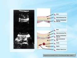
While waiting for the labs to return, a fluid challenge to complete and view his hypotension, you performed some tests with sonography and ultrasound, this is one part of the ultrasound, take a look at it and we will come back to it later but realize this is the view of the heart, left atrium, blood going through the mitral valve into the left ventricle, being pumped out into the aorta through the aortic valve and up here is the right ventricle, we will come back to this.
 RSS Feed