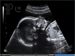
The EPSS is definitely greater than two, therefore, the ejection fraction is definitely less than thirty percent, now we are seeing part of a fast scan, the liver is noted to be more anterior, the right kidney is at the tip of this arrow and then Morrison’s pouch is the space in between, now this is a potential bedside sonography space and there is at the tip of the fat arrow, that’s free fluid, it doesn’t belong there, the CBC revealed a profound anaemia so medical decisions were made with these assumptions, this patient did not have a pulmonary embolism as shown by the CT angiography.

The new onset of congestive heart failure CHF could be quantified with an ejection fraction that was significantly decreased at less than thirty percent, the anaemia was probably from an inter abdominal bleed, the fast scan showed free fluid and the patient had a very significant anaemia, the bleed might very well be from his DIC and lastly the CHF possibly could be due to askemia from the acute bleed, obviously this would change your clinical management, whether to call surgery for the abdominal bleed, how much fluid to give, many decisions could be made based upon this ultrasound, based upon both ultrasounds so see the next presentation of test your sonography skills to assess your ability to interpret and utilize bedside sonography or see one of the vignettes that we have prepared for you.

 RSS Feed